产品推荐
细胞生物学
酶免试剂盒
细胞因子(重组蛋白)
生化试剂
实验用抗体
分子生物学
蛋白研究
实验室仪器
实验室耗材
技术服务
联系我们
上海研卉生物科技有限公司
地址:上海市嘉定区叶城路1288号
电话:021-69000211
联系人:李先生
手机:18702156455
传真:021-69000211
MSN:yanhuibio@hotmail.com
QQ:1320534253,245676794
邮箱:yanhuibio@163.com
线粒体自噬检测试剂盒
货 号:MD01
产品规格:盒
原 产 地:同仁
参考价格:4580 (参考价格,以实际价格为准)
优惠价格:3980
产品详细信息
线粒体自噬检测试剂盒 Mitophagy Detection Kit
货号:MD01

线粒体(mitochondria)是细胞内制造能量的场所,在细胞器中扮演着重要的角色。近年有报告指出,去极化线粒体的积累引起的阿尔茨海默病(Alzheimer‘s
disease)与帕金森病(Parkinson’s
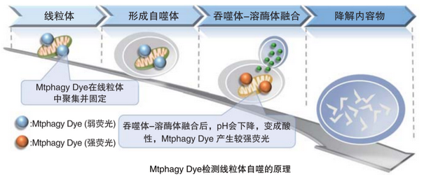
disease),可能与线粒体自噬有关。通过线粒体自噬(mitophagy)系统,将氧化应激、DNA损伤因素导致的去极化线粒体,隔离包裹成自噬体(autophagosome)后、再与溶酶体(lysosome)融合后降解。
本试剂盒包括“线粒体自噬体染料(Mtphagy
Dye)和溶酶体染料(Lyso
Dye)。MtphagyDye通过化学键结合在细胞内的线粒体上,保持较弱的荧光。通过诱导线粒体自噬,线粒体自噬体会与溶酶体融合,Mtphagy
Dye的荧光强度会增大。通过本试剂盒包含的Lyso Dye,单独标记溶酶体,来确认这一融合过程。
〇Mitophagy Induction and Detection of Mitochondrial Membrane Potential Changes
Mitochondrial condition in the
carbonyl cyanide m-chlorophenyl hydrazine (CCCP)treated
Parkin-expressing HeLa cells was compared with untreated cells using
Mitophagy Detection Kit (MD01) and JC-1 MitoMP Detection Kit (MT09).
〇 Result
Mitophagy was not detected in
untreated cells and the membrane potential was normal. However,
reduction of membrane potential and mitophagy were observed in treated
cells.

〇Detecting Condition
Mitophagy Detection]
Ex: 561 nm, Em: 570-700 nm
[Mitochondrial Membrane Potential Detection]
Green Ex: 488 nm, Em: 500-550 nm
Red Ex: 561 nm, Em: 560-610 nm
〇Experimental Condition
Transfection of Parkin plasmid to HeLa cells
HileyMax (H357) was used to
transfect Parkin plasmid to HeLa cells (Parkin plasmid/HilyMax reagent:
0.1μg/0.2 μL) by incubating overnight.
Detection of Mitophagy
1. Add 0.1 μmol/L Mtphagy working solution to Parkin expressing HeLa cells and incubated for 30 minutes at 37 ℃
2. Wash cells with HBSS
3. Add 10 μg/mL CCCP/MEM solution and inclubate for 2 hours at 37 ℃
4. Observe under fluorescence microscope
Detection of Mitochondrial Membrane Potential
1. Add 10 μg/mL CCCP/MEM solution to Parkin expressing HeLa cells and incubate for 1.5 hours at 37 ℃
2. Add 4 μmol/L JC-1 working solution (final concentration: 2 μmol/L) and incubate for 30 minutes at 37 ℃
3. Wash with HBSS and add Imaging Buffer Solution.
4. Observe under fluorescence microscope
Q:对比原有方法有什么优势?
		A:与使用pH敏感mt-Keima蛋白法比较,本试剂盒使用小分子荧光探针,不需要转染荧光蛋白。


		与一般活细胞成像的荧光探针一样染色观察即可。
Q:Mtphagy Dye和Lyso Dye的储存液可以保存多久?
A:Mtphagy Dye和Lyso Dye的储存液在-20℃可以稳定保存1个月。建议按照用量分装保存。
Q:Mtphagy Dye和Lyso Dye的工作液可以保存多久?
A:现配现用。
Q:培养基中的酚红影响检测吗?
A:如果是使用共聚焦显微镜的话几乎没有影响,如果使用落射荧光显微镜观察背景会高
(参考如下图片)。
因此使用落射荧光显微镜时,Working solution染色时要使用不含酚红的培养基或HBSS。
图片翻译:落射荧光显微镜、共聚焦显微镜
落射荧光显微镜 共聚焦显微镜

Q:荧光显微镜的推荐波长是什么?
A:各试剂分别推荐波长如下。
Mtphagy Dye ∶Ex:500~560 nm,Em:670~730 nm
Lyso Dye ∶Ex:350~450 nm,Em:500~560 nm
说明书中的「激发/发射光谱」和「荧光显微镜的检测例」参考如下。